Diagnosis Of Tuberculosis: Past, Present & Future
- March 18,2022
- 2 Min Read

Tuberculosis (TB) remains one of the leading infectious diseases throughout the world. The disease is caused by a bacterium called Mycobacterium tuberculosis (MTB). The bacteria usually attack the lungs but can infect any part of the body. If not treated properly, TB can be fatal. Early detection and appropriate treatment of infectious cases is the most important control strategy for TB.(1)
CLINICAL DIAGNOSIS
Clinical signs and symptoms develop only in 5–10% of the infected individuals. Tuberculosis primarily affects the lower respiratory system and these patients usually present with pulmonary disease. Prominent symptoms for pulmonary TB are chronic productive cough, hemoptysis, low-grade fever, night sweats, loss of appetite, malaise, fatigue, and weight loss. Tuberculosis may also affect extrapulmonary organs and manifest as lymphadenitis; kidney, bone, or joint involvement; meningitis; or disseminated (miliary) disease. Symptoms of extra-pulmonary TB depend on the organs involved.(2)
RADIOGRAPHY
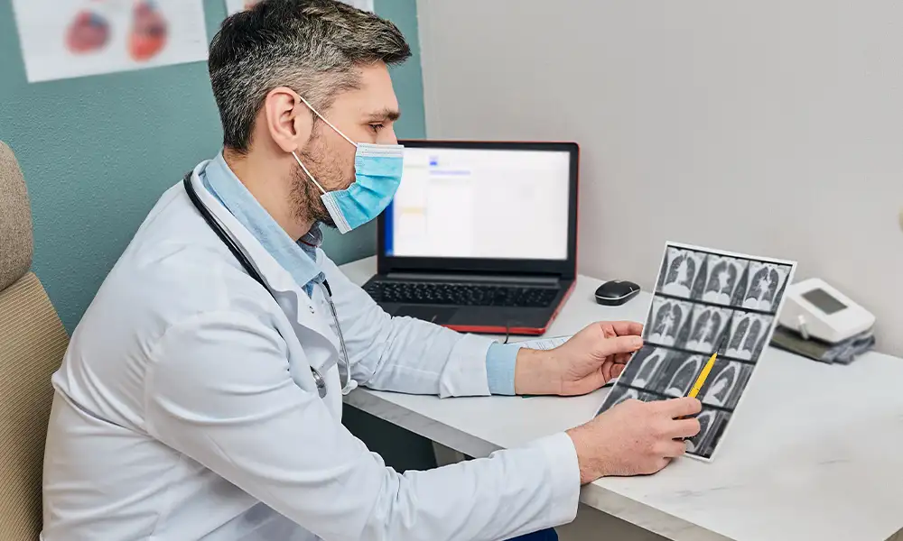
Chest X-ray (CXR) is a screening tool and are used as a diagnostic aid to differentiate between primary and secondary tuberculosis. Primary disease is usually characterized by a single lesion in the middle or lower right lobe with enlargement of the draining lymph nodes. Endogenous reactivation is often accompanied by a single lesion (cavitary) in the apical region, with unremarkable lymph nodes and multiple secondary tubercles. Miliary lesions, which are small granulomas, resemble millet seeds spread throughout the lung fields. Chest X-ray can help in the detection of pulmonary TB but do not provide any etiological diagnosis. It also can’t detect latent infection.(2) More powerful imaging techniques such as computerized tomography (CT) can help in the diagnosis of cases wherein X-ray findings are nebulous or equivocal.
SMEAR MICROSCOPY
Microscopic evaluation of stained smears made from sputum, urine or aspirated body fluids has been a rapid and inexpensive screening method for mycobacteria within clinical specimens for over 100 years and even today, in resource-poor settings, the diagnosis of TB relies on Ziehl-Neelsen smear microscopy with the light microscope.
Smear microscopy with the light microscope is a relatively insensitive methodology for the diagnosis of TB and only detects about 60-70% of the TB cases.(1)
An alternative for the light microscope is the fluorescence microscope. Fluorescent microscopy of MTB is reportedly more sensitive as compared to light microscopes and reduces the screening time by up to 25% as it allows visualization at a lower magnification.(1)
The World Health Organization (WHO) has endorsed the global phaseout of conventional Ziehl-Neelsen light microscopy in favour of Fluorescent AFB staining.(1)
CULTURE-BASED METHODS FOR TB DIAGNOSIS
The gold standard test for the diagnosis of TB is the isolation of MTB on a culture medium.
- Solid Culture Media:
Until the early nineties culturing was usually done on solid egg-based media like Lowenstein Jensen (LJ medium). A drawback of culturing on these solid media is that MTB is a slow-growing organism with a generation time of 14–15 hours and the colonies appear only in about two weeks and sometimes may be delayed up to 6–8 weeks.(2) Hence, cultures are typically held for 6-8 weeks before being discarded and reported as negative.(1)
- Culture in Broth:
Mycobacteria grow faster in broth than on solid media. Growth of MTB from clinical specimens takes an average of 10 days by broth systems versus 20-25 days on solid media. A drawback of this method is the contamination rate of liquid medium, which seems to be higher in comparison to the solid media. To overcome contamination with other microorganisms, some of the commercially available broth media are supplemented with an antibiotic mixture.(1)
There are 3 FDA approved platforms for the semi-automated broth-based culture of mycobacteria(1):
-
BACTEC MGIT 960 system (Becton Dickinson Microbiology Systems)
-
VersaTREK system (Trek Diagnostic Systems)
-
MB/BacT Alert 3D (bioMérieux)
DNA BASED TESTS FOR TB DIAGNOSIS
- Classic nucleic acid amplification tests (NAATs):
Several methodologies have been published for the detection of MTB using the polymerase chain reaction (PCR) assay, using primers to amplify a specific DNA fragment. These NAATs can give results in 3-6 hours.(1)
Although NAATs are rapid and have demonstrated excellent specificity, their performance characteristics can vary and their sensitivity still does not equal that of culture-based methods, especially for smear-negative samples.(1)
- Loop-mediated isothermal amplification test (LAMP):
This technology amplifies target DNA under isothermal conditions (about 65°C) and is designed to visually detect DNA directly from clinical samples, in less than two hours and with minimal instrumentation. Following the review of the latest evidences, WHO recommends that TB-LAMP can be used as a replacement for smear microscopy in adults with signs and symptoms of TB and can also be considered as a follow-on test to microscopy in adults with signs and symptoms of pulmonary TB, especially when further testing of sputum smear-negative specimens is necessary.(1)
- Xpert MTB/RIF nucleic acid amplification assay:
Xpert MTB/RIF is an automated, cartridge-based NAAT for simultaneous detection of TB and rifampicin resistance directly from sputum in less than two hours. A diagnostic test accuracy review including 27 unique studies concluded that, in comparison with smear microscopy, Xpert MTB/RIF increased TB detection among culture-confirmed cases by 23%. MTB/RIF pooled sensitivity was 95% and pooled specificity was 98%.(1)
Xpert MTB/RIF is rapidly being replaced by the more sensitive Xpert Ultra.(1) Compared to previous generations, Ultra test cartridges have a larger chamber for DNA 106 amplification than Xpert MTB/RIF, and two multicopy amplification targets for TB, IS6110 and 107 IS1081, lower limit of detection of 16 colony forming units (CFU)/ml.(3)
- Truenat MTB and Truenat MTB plus:
Truenat MTB and Truenat MTB plus is a new chip-based, micro real-time polymerase chain reaction (PCR) test that detects tubercle bacilli in sputum samples in approximately one hour. The overall sensitivity of the Truenat MTB assay is 83% and that of the MTBPlus assay is 89%. Specificity is 99% for the MTB and 98% for the MTBPlus assay.(1)
SERODIAGNOSIS OF TB
An antibody detection based diagnostic test in a user-friendly format could potentially replace microscopy and extend tuberculosis diagnosis to lower levels of health services. However, following recent meta-analyses and systematic reviews, WHO currently recommends against their use for the diagnosis of pulmonary and extrapulmonary TB.(1)
DIAGNOSIS OF LATENT TB
Persons with latent TB infection (LTBI) are infected with MTB but do not have TB disease. For over a century, the tuberculin skin test (TST), or Mantoux test was the only screening tool for latent TB. Although the Mantoux test is an inexpensive diagnostic tool, the test has a low specificity in patients with a previous history of BCG vaccination or exposure to nontuberculous mycobacteria (NTM).(1)
- Interferon-γ release assays:
Interferon-γ release assays (IGRAs) were developed to address the shortcomings of the Mantoux test. These in vitro blood tests assess the immunologic reaction of cytokines (interferon- γ) to antigens ESAT-6 and CFP-10, specific to M. tuberculosis.(1)
There are two commercially available IGRAs(1):
-
Quantiferon TB Gold
-
T-SPOT
Several published studies have demonstrated better performance of these tests over the Mantoux tests in the diagnosis of a TB infection. Despite these studies, the lack of a reference standard test for LTBI makes it difficult to access the true accuracies of these assays.(1)
Systematic reviews have suggested that IGRA performance differs in high-versus low TB and HIV incidence stings, with relatively lower sensitivity in high-burden settings. Consequently, WHO recommends that IGRAs and Mantoux should not be used for the diagnosis of pulmonary or extra-pulmonary tuberculosis in low- and middle-income countries.(1)
MYCOBACTERIUM SPECIES IDENTIFICATION
The genus Mycobacterium comprises more than 150 species and several newly discovered pathogenic nontuberculous mycobacterial (NTM) species. The NTM species most frequently associated with pulmonary disease are Mycobacterium avium, Mycobacterium kansasii, and Mycobacterium abscessus and in some countries, pulmonary infection with NTM has become more important than TB.(1)
Historically, mycobacteria were identified by phenotypic methods, such as morphological characteristics, growth rates, preferred growth temperature, pigmentation and a series of biochemical tests for instance reduction of nitrates, catalase, niacin accumulation, urease activity, Tween 80 hydrolysis, arylsulfatase, urease activity and iron uptake. These tests are laborious, time-consuming and in certain instances misidentification may occur because different species may have indistinguishable morphological and biochemical profiles.(1)
There are several molecular technologies that are currently employed by clinical diagnostic laboratories to identify isolates from culture including(1):
-
Nucleic acid hybridization probes
-
Line probe hybridization assays
-
Matrix-assisted laser desorption/ionization time-of-flight mass spectroscopy (MALDI-TOF MS)
-
DNA sequencing
-
Immunochromatography
THE FUTURE OF TB DIAGNOSIS – WHOLE GENOME SEQUENCING
Whole genome sequencing (WGS), the next-generation sequencing (NGS) based technology allows the high throughput sequence read of the entire genome of a microorganism.(2)
Whole genome sequencing poses an advantage to ideally detect all the mutations with their functional categorization as compared to Xpert MTB/RIF and line probe assays which are limited to identifying mutations in specific locus and are unable to distinguish whether a mutation leads to a change in amino acid sequence. Furthermore, WGS can identify the resistance determining mutations in new drugs, for example, bedaquiline and delamanid.(2)
The WHO is soon likely to publish a technical guideline addressing the application and interpretation of DNA sequencing technology in TB diagnostics.(2)
RECOMMENDED DIAGNOSTIC MODALITIES
Xpert MTB/RIF or Xpert Ultra MTB/RIF should be used as an initial diagnostic test for TB, rather than smear microscopy/culture methods.
In the 2021 update to its TB diagnosis guidelines, the WHO recommends the Xpert platform be used for diagnosis of pulmonary, extrapulmonary and disseminated TB.(4)
In case of a negative result on Xpert Ultra, broth-based culture can be done if there still exists a strong clinical suspicion of TB. (4)
References:
1. Chopra KK, Sidiq Z, Hanif M, Dwivedi KK. Advances in the diagnosis of tuberculosis- Journey from smear microscopy to whole genome sequencing. Indian J Tuberc. 2020 Dec;67(4):S61–8.
2. Acharya B, Acharya A, Gautam S, Ghimire SP, Mishra G, Parajuli N, et al. Advances in diagnosis of Tuberculosis: an update into molecular diagnosis of Mycobacterium tuberculosis. Mol Biol Rep. 2020 May;47(5):4065–75.
3. MacLean E, Kohli M, Weber SF, Suresh A, Schumacher SG, Denkinger CM, et al. Advances in Molecular Diagnosis of Tuberculosis. Suzanne Kraft C, editor. J Clin Microbiol. 2020 Sep 22;58(10):e01582-19.
4. World Health Organization. WHO consolidated guidelines on tuberculosis: module 3: diagnosis: rapid diagnostics for tuberculosis detection [Internet]. 2021 update. Geneva: World Health Organization; 2021 [cited 2022 Mar 16]. Available from: https://apps.who.int/iris/handle/10665/342331
Want to book a test? Fill up the details & get a callback
Most Viewed
Premarital Health Screening
- 20 Min Read
Typhoid - Signs and Symptoms
- 3 Min Read
Home Isolation Guidelines - Covid-19 Care
- 5 Min Read
HLA B27 Detection: Flow Cytometry & PCR
- 1 Min Read


